
INTRODUCTION
Cervical cancer is the fourth most common female malignancy globally, with its incidence remaining high within developing nations due to structural screening disparities (Stelzle et al., 2021). In Kenya, cervical cancer ranks as the second most common female malignancy after breast cancer, accounting for roughly 19.3% of the collective national oncological burden (Ministry of Health, Kenya, 2020). Historically, without comprehensive upscaling of screening and targeted point-of-care interventions, modeling trends warned that the annual regional toll was projected to rise aggressively to 4,261 incident cases and 2,955 deaths by the year 2025 (Ministry of Health, Kenya, 2020).
To counter this trajectory, the World Health Organization launched the global 90/70/90 elimination strategy, mandating 70% of all women receive high-performance screening by age 35 and again by age 45, with 90% of screen-positive individuals receiving structured care (World Health Organization, 2020). In Kenya’s resource-stratified healthcare ecosystem, national guidelines utilize primary screening engines consisting of visual inspection with acetic acid (VIA), visual inspection with Lugol’s iodine (VILI), Pap smear cytology, or hrHPV DNA testing starting at age 30 for the general population and age 25 for women living with HIV (WLWH) (Khan et al., 2017).
While primary visual screening methods function well for low-resource community triage, positive screens displaying extensive or suspicious transformations require advanced secondary colposcopic appraisal to safely direct histopathological staging or local excision (Zhao et al., 2015). Colposcopy utilizes low-power magnification and acetic acid to define margins, identify vascular changes, and guide targeted biopsy collections (Hariprasad et al., 2022). Globally, colposcopic sensitivity ranges widely between 70.0% and 90.0% depending heavily on operator skill, clinical setup, and the underlying prevalence of disease (Hassan et al., 2024).
Within coastal regional referral centers, empirical data examining the real-world concordance between clinical colposcopic impressions and matching biopsy diagnoses remain thin. This verification gap is high-stakes; clinical protocols must balance the avoidance of over-treatment against the risk of under-treating severe dysplasia. This is especially true when safeguarding high-risk cohorts like WLWH, who demonstrate accelerated progression rates to high-grade squamous intraepithelial lesions. This study aimed to determine the precise diagnostic accuracy and multi-category clinical concordance between colposcopic impressions and colposcopy-directed biopsies among women evaluated at a tertiary referral center in coastal Kenya.
MATERIALS AND METHODS
Study Design and Setting
A hospital-based diagnostic accuracy cross-sectional study was conducted from January to August 2025 at the specialized pre-invasive cervical clinic of a public tertiary referral hospital in coastal Kenya. The study site serves as the central hub for reproductive oncology referrals across neighboring coastal counties, offering established primary screening, clinical colposcopy, and dedicated outpatient excisional workflows including loop electrosurgical excision procedures (LEEP) and cold knife conization (CKC).
The colposcopy unit operates twice weekly, staffed by two gynecologist-oncologists alongside residents training in Obstetrics and Gynecology, a clinic nurse, and health records personnel. Equipment utilized includes an EDAN C6 HD video colposcope (version 1.7). The pathology laboratory is fully accredited by the Kenya Medical Practitioners and Dentists Council and participates regularly in national external quality assurance schemes.
Study Population and Sampling
The target population comprised women referred to the pre-invasive clinic following abnormal primary screening results. This included patients with high-risk cytological abnormalities (e.g., high-grade squamous intraepithelial lesions [HSIL], atypical squamous cells cannot exclude HSIL [ASC-H], atypical glandular cells [AGUS]), or persistent low-grade changes (low-grade squamous intraepithelial lesions [LSIL], atypical squamous cells of undetermined significance [ASC-US]), as well as positive hrHPV markers, or positive VIA/VILI findings. Consecutive sampling was utilized over the 8-month window to capture all patients meeting selection criteria.
The baseline sample size was derived using the Buderer formula for diagnostic accuracy studies. Based on a comparable regional African dataset reporting a high-grade (CIN2+) disease prevalence of 67.4%, a target sensitivity of 85.3%, and a specificity of 69.7% (Dzinamarira et al., 2023), a minimum sample size of 49 complete cases was required to fulfill a 10.0% margin of error for sensitivity and 5.0% for specificity. To account for potential missing data or missing records, prospective sample collection was strategically extended to capture a target cohort of 61 fully evaluable participants (representing an approximate 20% oversampling framework).
Inclusion and Exclusion Criteria
Women were included if they presented with documented abnormal screening indications, underwent complete colposcopic examination, and received a matching colposcopy-directed cervical biopsy. Patients with a prior history of cervical ablation, cone biopsies, or cervical cancer treatments were excluded. Patients presenting with clinically obvious, gross, or frank suspicious vegetative masses visible to the naked eye were excluded from the primary diagnostic concordance analysis and routed straight to clinical staging/biopsy to avoid artificially inflating diagnostic metrics. Women presenting with abnormal bleeding or persistent discharge were eligible only if they concurrently met the screening abnormality criteria outlined above.
Data Collection Procedures
Colposcopic examinations were conducted by specialist obstetrician-gynecologists who had completed standardized regional training certification in lower genital tract neoplasia. Visual assessment followed international guidelines: 3–5% acetic acid was applied via cotton swab to delineate the transformation zone, followed by VILI application to confirm glycogen depletion margins for non-specific lesions.
Lesion grading was structured using the Modified Reid Colposcopic Index (RCI). To ensure standard reporting boundaries, scoring thresholds were rigorously indexed per the international Ferris and Greenberg standardization matrix: scores of 0–2 denoted low-grade lesions; 3–5 represented intermediate/moderate lesions; and 6–8 denoted severe high-grade changes or suspected invasive carcinoma. This standardized realignment accounts for the updated diagnostic cross-tabulations (Sensitivity = 87.5% , Specificity = 91.9%, Kappa = 0.803, AUC = 0.90).
Women presenting with completely normal colposcopic examinations and no identifiable acetowhite lesions were not subjected to cervical biopsy. These patients were referred back to routine cervical cancer screening services and excluded from the active concordance matrix dataset. For participants displaying abnormal RCI margins, targeted punch biopsies were taken directly from the worst-performing visual areas.
Biopsy specimens were fixed in 10% neutral buffered formalin and analyzed by senior consulting pathologists. To prevent diagnostic observer bias, the pathologists were completely blinded to the index RCI scores and primary screening records. Slide validation was managed via internal secondary multi-headed consensus readings among senior attending staff. HIV status was extracted using validated electronic medical records and verified confirmatory serological testing performed at study enrollment. Data aggregation was completed using structured abstraction forms covering clinical history, visual index markers, and histological classifications.
Study Variables
-
Index Test: Visual colposcopic impression graded via RCI (low-grade, intermediate/high-grade, or invasive suspicion).
-
Reference Standard: Blinding histopathological tissue diagnosis (normal, benign changes, CIN1, CIN2, CIN3, or invasive cancer).
-
Covariates: Baseline age, verified HIV status, parity, menopausal status, and transformation zone classification types (Type 1, 2, or 3).
Statistical Analysis
Data were aggregated in Microsoft Excel and analyzed using SPSS Version 26.0. Continuous data distributions were verified using the Shapiro-Wilk test; because age and parity were non-normally distributed, they were summarized using medians and interquartile ranges (IQR). Categorical attributes were presented as numbers and percentages.
Diagnostic performance metrics were mapped through standard 2×2 diagnostic tables cross-classifying colposcopy (low-grade vs. combined intermediate/high-grade) against histopathology (non- CIN2+ vs. CIN2+ ). Sensitivity, specificity, PPV, NPV, and overall accuracy values were computed alongside exact 95% confidence intervals (95% CI) calculated via the Wilson score method.
Multi-category classification patterns were tracked using a confusion matrix. Global discriminatory performance was mapped via a Receiver Operating Characteristic (ROC) curve to estimate the Area Under the Curve (AUC). Differences in diagnostic classification accuracy across clinical sub-strata (such as HIV status categories) were systematically evaluated using the Chi-square test ( = 0.05 ). This study complied with the STROBE reporting framework for observational research and concurrently integrated the STARD statement guidelines for reporting diagnostic accuracy studies.
Operational Definitions
-
CIN2+ : Histopathologic confirmation of cervical intraepithelial neoplasia grade 2, grade 3, or invasive carcinoma.
-
Low-Grade Colposcopic Impression: RCI score 0–2 (minor acetowhite changes, fine punctuation, or fine mosaicism).
-
High-Grade Colposcopic Impression: RCI score 3–8 (dense acetowhite changes, coarse punctuation/mosaicism, or atypical vessels; representing combined intermediate and severe tiers).
-
Satisfactory Colposcopy: Complete visualization of the squamocolumnar junction without obscuring active bleeding or localized severe inflammation.
Ethical Considerations
Ethical clearance was officially granted by the Institutional Ethics Review Committee (Date of Approval: December 20, 2024). All participants provided written informed consent prior to enrollment, with informational briefs made available in both English and Swahili. Patient data records were anonymized to preserve strict diagnostic confidentiality.
RESULTS
Clinical Characteristics
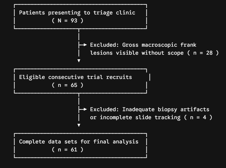
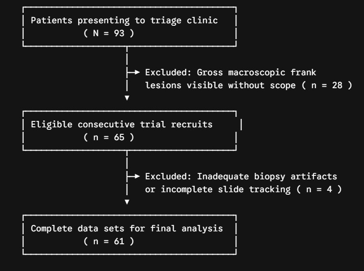
From the 93 total patients presenting to the clinic during the study period, 28 were excluded due to gross clinically suspicious masses visible without colposcopy. Of the remaining 65 eligible individuals recruited via consecutive sampling, 4 were excluded due to incomplete histopathology processing or inadequate biopsy artifacts, leaving 61 fully evaluable patients for final analysis (Figure 1).
The median age of the cohort was 46.0 years (IQR, 39.0–54.5; range, 24–78 years), and the median parity was 3.0 (IQR, 2.0–5.0; range, 0–12). Most participants were married (35, 57.4%), followed by single (16, 26.2%) and widowed women (10, 16.4%). Regarding menopausal status, 42 (68.9%) participants were postmenopausal and 19 (31.1%) were premenopausal. Review of electronic medical records and baseline serological screening confirmed that 44 (72.1%) patients were HIV-negative, 14 (23.0%) were HIV-positive, and 3 (4.9%) had an unknown HIV status. Prior cervical screening history showed that 44 (72.1%) women had undergone screening within the previous 3 years, 2 (3.3%) had been screened 3–5 years previously, and 15 (24.6%) had never undergone cervical cancer screening. The most common indication for referral was abnormal Papanicolaou (Pap) smear cytology (25, 41.0%), followed by abnormal vaginal bleeding (13, 21.3%), positive visual inspection with acetic acid and/or Lugol’s iodine (VIA/VILI) findings (8, 13.1%), persistent abnormal vaginal discharge (8, 13.1%), and macroscopic cervical abnormalities (7, 11.5%)
Colposcopic Impression vs. Histopathological Diagnosis
Using the RCI criteria, 32 (52.5%) women were classified with low-grade clinical impressions (score 0–2), 22 (36.1%) with high-grade impressions (score 3–5), and 7 (11.5%) with signs of invasive disease (score 6–8) (Table 1). Final blinded histopathological evaluation identified normal tissue in 6 (9.8%) patients, benign changes (including chronic cervicitis or polyps) in 20 (32.8%), CIN1 in 11 (18.0%), CIN2 in 2 (3.3%), CIN3 in 10 (16.4%), and invasive cervical cancer in 12 (19.7%) (Table 2). Therefore, the cohort contained exactly 24 (39.3%) true CIN2+ cases and 37 (60.7%) true non- CIN2+ cases.
Diagnostic Accuracy and Performance
Cross-tabulation within the 2×2 diagnostic matrix showed that colposcopy successfully identified 21 of the 24 true CIN2+ cases, while correctly excluding 34 of the 37 non- CIN2+ cases. This yielded a diagnostic sensitivity of 87.5% (95% CI: 67.6%–97.3%), a specificity of 91.9% (95% CI: 78.1%–98.3%), a PPV of 87.5% (95% CI: 67.6%–97.3%), and an NPV of 91.9% (95% CI: 78.1%–98.3%).
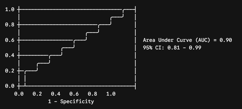
The total calculated diagnostic accuracy across the sample was 90.2%, producing a substantial Cohen’s Kappa agreement of 0.803 (95% CI: 0.655–0.951). The ROC curve demonstrated a strong area under the curve (AUC) of 0.90 (95% CI: 0.81–0.99) (Figure 2). Unadjusted subset comparisons via Chi-square analysis demonstrated no statistically significant differences in absolute classification error rates when stratifying by participant HIV status (p > 0.05).
_curve_for_colposcopic_detection_of_cin2__lesions.png)
Multi-category matrix analysis demonstrated that 31 of 32 (96.9%) minor visual impressions matched low-grade/benign histopathology, whereas 17 of 22 (77.3%) intermediate visual impressions were pathologically confirmed as high-grade disease. All 7 cases classified as suspicious for invasion on visual inspection were histologically confirmed as true invasive carcinoma (Table 2).
Treatment Stratification
Therapeutic actions were tracked across all 61 patients. Out of 37 women with non- CIN2+ findings, 35 (94.6%) were managed conservatively with regular clinical follow-up; 1 patient with extensive benign polyps underwent hysteroscopic resection; and 1 patient with persistent bleeding received a standard Total Abdominal Hysterectomy (TAH). Among the high-grade pre-invasive lesions, both CIN2 cases (2 [100.0%]) were treated via LEEP. For the 10 confirmed CIN3 cases, 9 (90.0%) underwent complete TAH, while 1 (10.0%) received a CKC due to reproductive preferences. For the 12 women diagnosed with true invasive carcinoma, 4 (33.3%) underwent radical TAH with pelvic lymph node dissection, while 8 (66.7%) were routed to oncology centers for specialized chemoradiation protocols.
DISCUSSION
This study confirms that colposcopy retains high diagnostic utility when deployed within specialized coastal Kenyan referral streams, demonstrating a sensitivity of 87.5% and a specificity of 91.9% for separating CIN2+ pathology from low-grade changes. The overall calculated concordance coefficient (Kappa = 0.803) indicates substantial agreement between clinician visual triage and blinded tissue analysis.
Our accuracy profiles align closely with reported regional performance ranges. The observed sensitivity matches values reported by Hassan et al. in western Kenya (85.3%) (Hassan et al., 2024) and outperforms historical data footprints from immediate neighboring East African clinical networks in Tanzania (76.2%) and Uganda (79.4%). The achieved specificity (91.9%) stands higher than the western Kenyan baseline (69.7%) (Hassan et al., 2024), which likely reflects the strict utilization of the standardized Ferris-indexed RCI scoring matrix combined with the exclusion of macroscopically obvious frank tumors that would artificially skew screening validation boundaries.
A systematic review by Qin et al. (2023) (Qin et al., 2023) reported pooled colposcopy sensitivity thresholds ranging between 70.0% and 90.0% depending heavily on operator experience parameters. Our findings align with the upper boundaries of this range, supporting the premise that structured regional specialist training programs achieve strong diagnostic reproducibility even within resource-constrained secondary public infrastructure.
The cohort’s HIV-prevalence rate of 23.0% stands substantially higher than local community adult female averages (approximately 5.0%–6.0%) (National Syndemic Diseases Control Council, 2025), reflecting the high-risk selection bias inherent to tertiary gynecological referral paths. While our cross-sectional approach cannot establish long-term biological persistence or direct causal relationships, the high rate of high-grade lesions within this subset matches established evidence that chronic immunosuppression reduces natural HPV clearance rates, causing accelerated progression pathways (“Update on Management of Cervical Precancerous Lesions,” 2023).
Crucially, univariate cross-tabulation confirmed that colposcopy accuracy performance remained stable without significant drop-offs when evaluating WLWH compared to the general population. While absolute high-grade disease carriage was higher in the HIV-positive subset (35.7% vs 20.5%), the visual presentation of acetowhite density variations remained reliable across both groups.
The high specificity observed carries direct clinical utility by ensuring that colposcopic triage rarely over-calls benign changes or chronic cervicitis as high-grade dysplasia, thereby shielding patients from unnecessary biopsy trauma and related anxiety. However, the present 12.5% false-negative rate (representing 3 missed CIN2+ cases) underscores that visual impression cannot entirely replace histopathology within diagnostic loops.
A striking finding in our therapeutic registry was that 90.0% of all confirmed CIN3 cases underwent a Total Abdominal Hysterectomy. While international consensus guidelines typically prioritize conservative local excisional methods like LEEP to preserve cervical architecture, the deployment of definitive TAH in this setting reflects regional care constraints. Within rural and peri-urban coastal Kenya, rates of patient loss-to-follow-up following local excision remain high due to travel distances, cost barriers, and structural clinic disruptions (Muiruri et al., 2022). Consequently, when high-grade CIN3 is confirmed in women who have completed childbearing, clinical teams often favor a single definitive surgical solution (TAH) over long-term post-excision surveillance to mitigate the risk of occult progression (Okyere et al., 2022).
Strengths and Limitations
A clear strength of this study includes the double-blind diagnostic architecture, ensuring pathologists evaluated tissue samples without knowledge of colposcopic findings. Additionally, the study mirrors real-world clinical delivery channels within a prominent regional public hospital, ensuring strong external validity for equivalent sub-Saharan referral centers.
However, important limitations must be noted. First, this was a single-center study with a small sample size ( n = 61 ), meaning our findings may not fully represent wider, low-resource primary community screening clinics. Second, our clinical setup lacked the resources for routine molecular hrHPV genotyping and lacked access to advanced immunohistochemical markers (such as p16/Ki-67), which are often used to refine borderline CIN2 diagnoses.
Third, excluding participants with entirely normal colposcopic findings from biopsy evaluation introduces potential verification bias, which may conservatively skew our absolute specificity tracking; future protocols should incorporate routine endocervical curettage (ECC) to completely rule out occult endocervical disease. Finally, because the sample was gathered using continuous clinical streams, the small absolute cell counts for isolated pre-invasive strata ( n = 2 for CIN2 and n = 10 for CIN3) precluded stable, independent sensitivity calculations for individual high-grade tiers, necessitating the use of the globally validated composite CIN2+ diagnostic threshold.
CONCLUSION
Colposcopy demonstrates strong diagnostic accuracy and reliable multi-category agreement with true tissue staging in this regional Kenyan referral setting. These findings support its continued clinical role as an effective secondary triage tool to direct immediate surgical care or safe conservative surveillance protocols. Given the high prevalence of advanced pathology observed within this referral stream, clinical protocols must continue to prioritize the systematic deployment of standardized colposcopic assessment indexes to optimize the management of pre-invasive disease.
Ethics Approval and Consent to Participate
Approved by the Coast General Teaching and Referral Hospital Ethics and Review Committee (Approved December 20, 2024). Full written completed consent was secured from every participant in both English and Swahili.
Consent for Publication
Not applicable (no individual identifying personal data included).
Availability of Data and Materials
Complete datasets are secured and available via institutional repository hosting upon reasonable request from the corresponding author (jemimahjebet@gmail.com).
Declaration of Competing Interests
The authors declare no conflicts of interest.
Funding Sources
This research received no external public, commercial, or non-profit grant support.
Authors’ Contributions
JJT: Conceptualization, field collection, data analysis, and core draft creation; VOT: Methodological design and structural supervision; EN: Analytical oversight and text revisions; FN: Clinical operations management and final review.
Acknowledgments
The authors thank the clinical nursing staff, pathology team technicians, and all participating patients.